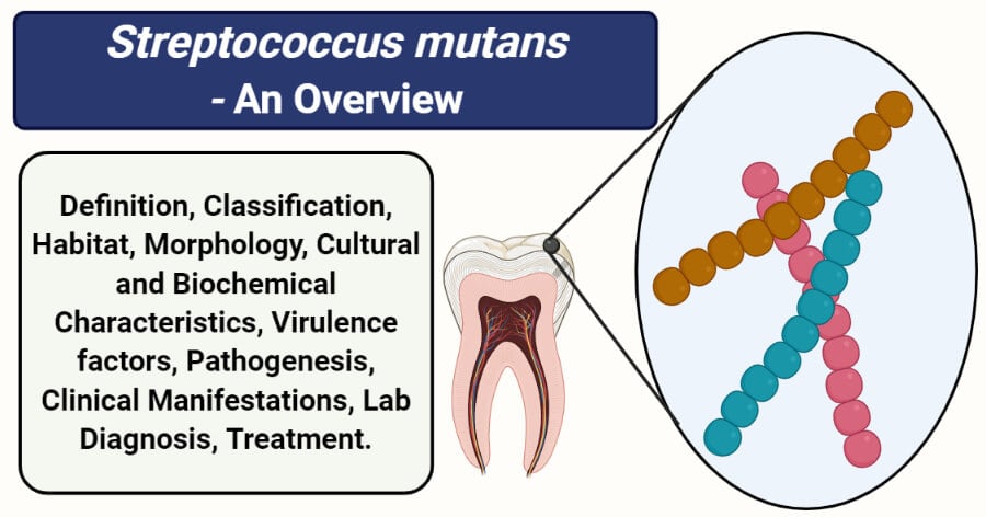
Streptococcus mutans is a Gram-positive coccus that is a major inhabitant of the oral cavity and is considered a significant contributor of tooth decay and cavities.
- Streptococci are spherical or ovoid cells, arranged in chains or pairs where many species are members of the commensal microflora on mucosal membranes of humans or animals, and some are highly pathogenic.
- S. mutans is a commensal present as a part of the human normal flora, mostly in the oral cavity. It is rarely pathogenic but might act as an opportunistic pathogen in some cases.
- It is also called a cariogenic bacterium as it is present in the oral cavity and in the multispecies biofilms on the surfaces of teeth and causes dental plaque and cavities.
- It is a dominant species with higher biomass in dental biofilms than other Streptococcus species, including S. sanguinis, S. mitis, and S. salivarius, due to its acid tolerance and thus the capability to live in low pH environment of oral cavities.
- S. mutans is one of the eight species belonging to the mutans group of Streptococci which are differentiated based on their heterogenicity in their genomic structure.
- It was first discovered and named by J Kilian Clarke in 1924 which were isolated and identified from carious lesions.
- Streptococcus mutans are major cariogenic organisms as a result of their ability to produce large quantities of glucans as well as acid, exceeding the buffering capacity of the oral environment.
Interesting Science Videos
Classification of Streptococcus mutans
- The genera Streptococcus belong to the lactic acid bacteria, which is a taxonomically diverse group of gram-positive, non-spore-forming cocci and rods defined by the formation of lactic acid as a sole or major endproduct of carbohydrate metabolism.
- Based on 16S rRNA gene sequence analysis the genus Streptococcus belongs within the low (< 50 mol%) G+C branch of the Gram-positive eubacteria, and is a member (type genus) of the family Streptococcaceae.
- The genus currently consists of over 50 recognized species which, for the most part, fall within “species groups” which are identified on the basis of different characteristics.
- S. mutans belongs to the mutans group because of its heterogeneous genomic structure.
The following is the taxonomical classification of S. mutans:
| Domain: | Bacteria |
| Phylum: | Firmicutes |
| Class: | Bacilli |
| Order: | Bacillales |
| Family: | Streptoococcaceae |
| Genus: | Streptococcus |
| Species: | S. mutans |
Habitat of Streptococcus mutans
- All the Streptococcus species are obligate parasites of mucosal membranes and, for some species, tooth surfaces of humans and several animals.
- Many of the species are life-long and dominant members of the commensal microflora on mucosal membranes of the upper respiratory tract, and some colonize the intestinal and genital tracts of humans and various animals.
- S. mutans however, exclusively colonize tooth surfaces and are present only after tooth eruption.
- Members of S. mutans may cause infection when introduced into normally sterile compartments of the body or in immunocompromised patients.
- The natural habitat of S. mutans is the human mouth, more specifically dental plaque, where the bacterium resides in multispecies biofilms that form on the surfaces of teeth.
- The oral cavity is a dynamic environment that undergoes large and rapid fluctuations in pH, nutrient availability and source, oxygen tension, temperature, and osmolality.
- The ability of S. mutans to survive under such an environment is attributed to its ability to produce acid as well as glucans from carbohydrates which enables the organism to maintain a favorable condition.
- The optimum temperature for the organism is the average body temperature of the host, but it is known to survive in a temperature range of 18-40°C.
Morphology of Streptococcus mutans

Figure: Gram-stained, thioglycollate broth culture specimen, revealed numerous, Gram-positive, Streptococcus mutans bacteria. Image Source: CDC/ Dr. Richard Facklam.
- The cells of S. mutans are coccoid, approximately 0.5–0.75 µm in diameter, but rod-shaped morphology may be evident on primary isolation from oral specimens.
- The arrangement of cells in S. mutans is characteristic of all Streptococci as the cells are arranged in pairs or as short- to medium-length chains. This arrangement is due to the presence of successive division planes that are parallel to one another as in rod-shaped bacteria.
- The cell wall consists of the shape-forming peptidoglycan (murein), various carbohydrate structures including teichoic acids, and a number of proteins, which form an interwoven complex.
- As in other gram-positive cell walls, the peptidoglycan consists of multiple glycan chains that are cross-linked through short peptides, and the glycan moiety is composed of alternating b-1,4-linked units of N-acetylglucosamine and N-acetylmuramic acid.
- Rhamnose is the primary carbohydrate of the cell wall, and Glycine is the major amino acid.
- The cell membrane is a typical lipid-protein bilayer, composed mainly of phospholipids and proteins.
- S. mutans also produce an array of proteins associated with the cell wall and are generally exposed on the outer surface of the cell wall.
- Members of this species also have different adhesins on their surface that mediate binding to salivary glycoproteins and bacteria-derived salivary components.
- These adhesins recognize extracellular matrix and serum components, particularly fibronectin and plasminogen, as well as host and other microbial cells.
Cultural characteristics of Streptococcus mutans
- The growth of S. mutans on ordinary nutrient media is generally low in contrast to that of other Gram-positive species.
- Growth is more profuse on media enriched with blood, serum, or a fermentable carbohydrate.
- To avoid competition and to inhibit other Gram-positive organisms, Selective Strep Agar is used as a selective media.
- S. mutans is facultatively anaerobic; while most strains grow in air growth is optimum at 37°C under the anaerobic condition with some strains CO2-dependent.
- A few strains have been reported to grow at 45°C, but no growth occurs at 10°C.
1. Nutrient Agar
- White to grey colored colonies of an average size of 1 mm in diameter. The colonies were round with raised elevation and an entire margin.
- Growth is mostly poor and requires air with supplied carbon dioxide.
2. Sucrose Agar
- Growth on sucrose-containing agar typically produces rough, heaped colonies, about 1 mm in diameter. Some strains may form smooth or mucoid colonies.
- The soluble extracellular polysaccharide is formed that is visible as beads, droplets, or puddles of liquid on or surrounding the colonies.
- The polysaccharide will, in some of the strains, result in coherent and adherent colonies that may be difficult to sub-cultivate on to agar plates or into fluid media.
3. Blood Agar
- Growth on blood agar after incubation anaerobically for 2 d produces colonies that are white or grey, circular or irregular, 0.5–1.0 mm in diameter.
- Sometimes, however, hard colonies tending to adhere to the surface of the agar and slightly pitting into the agar surface might be observed.
- Hemolytic reaction on blood agar is usually α-hemolytic or non-hemolytic with very occasionally strains to give β-hemolysis.
Biochemical Characteristics of Streptococcus mutans
The biochemical characteristics of Streptococcus mutans can be tabulated as follows:
| S.N | Biochemical Characteristics | Streptococcus mutans |
| 1. | Capsule | No capsule |
| 2. | Shape | Cocci |
| 3. | Catalase | Negative (-) |
| 4. | Oxidase | Positive (+) |
| 5. | Citrate | Negative (-) |
| 6. | Methyl Red (MR) | Negative (-) |
| 7. | Voges Proskauer (VR) | Positive (+) |
| 8. | OF (Oxidative-Fermentative) | Facultative anaerobes |
| 9. | Coagulase | Negative (-) |
| 10. | DNase | Negative (-) |
| 11. | Clumping factor | Negative (-) |
| 12. | Gas | Negative (-) |
| 11. | H2O2 | Negative (-) |
| 12. | Hemolysis | α, β-hemolytic |
| 13. | Motility | Non-motile |
| 14. | Nitrate Reduction | Negative (-) |
| 15. | Gelatin Hydrolysis | Negative (-) |
| 16. | Pigment Production | Variable |
| 17. | Bile esculin test | Positive (+) |
| 18. | Bacitracin | The majority of strains are resistant. |
| 19. | Lancefield group | Non-groupable |
Fermentation
| S.N | Substrate | Streptococcus mutans |
| 1. | Glucose | Positive (+) |
| 2. | Fructose | Positive (+) |
| 3. | Galactose | Positive (+) |
| 4. | Lactose | Positive (+) |
| 5. | Maltose | Positive (+) |
| 6. | Mannitol | Positive (+) |
| 7. | Mannose | Positive (+) |
| 8. | Raffinose | Positive (+) |
| 9. | Ribose | Negative (-) |
| 10. | Sucrose | Positive (+) Extracellular polysaccharide (dextran) is produced from sucrose. |
| 11. | Starch | Positive (+) |
| 12. | Trehalose | Positive (+) |
| 13. | Xylose | Negative (-) |
| 14. | Salicin | Positive (-) |
| 15. | Glycerol | Positive (+) |
| 16. | Dulcitol | Negative (-) |
| 17. | Cellobiose | Positive (+) |
| 18. | Rhamnose | Negative (-) |
| 19. | Arabinose | Negative (-) |
| 20. | Inulin | Positive (+) |
| 21. | Sorbitol | Positive (+) |
| 22. | Pyruvate | Negative (-) |
| 23. | Glycogen | Negative (-) |
Enzymatic Reactions
| S.N | Enzymes | Streptococcus mutans |
| 1. | Acetoin | Positive (+) |
| 2. | Acid Phosphatase | Positive (+) |
| 3. | Alkaline Phosphatase | Negative (-) |
| 4. | Ornithine Decarboxylase | Not determined |
| 5. | Hyaluronidase | Negative (-) |
| 6. | β-D-galactosidase | Negative (-) |
| 7. | Arginine Dehydrolase | Negative (-) |
| 8. | Neuraminidase | Negative (-) |
- S. mutans can hydrolyze arginine but cannot hydrolyze esculin and gelatin.
- They can tolerate 6.5% NaCl but cannot tolerate higher concentrations than that.
Virulence factors of Streptococcus mutans
Even though S. mutans hasn’t yet evolved to cause diseases, it is known to cause infections of the teeth and gums in immune-compromised patients. As one of the many etiological factors of dental caries, S. mutans is able to acquire new characteristics, allowing for the increased pathogenicity in specific environmental conditions. Beginning with the attachment to a solid surface, S. mutans is then capable of colonizing the oral cavity and also forming a bacterial biofilm. Other properties assisting S. mutans in colonizing the oral cavity include specific interaction with other microorganisms colonizing the oral ecosystem and the ability to survive in an acidic environment.
The following are some virulence factors of S. mutans in detail:
1. Biofilm
- Biofilms play a significant causative role in most oral cavity infections like dental caries.
- Biofilms present in the oral cavity are mostly proteinaceous structures, embedded in an exopolysaccharide matrix, consisting of bacterial cells anchored to solid surfaces, mostly the tooth enamel, tooth roots, or dental implants.
- The exact structure and composition of the exopolysaccharide matrix present on the outside of the biofilm is determined by the conditions of the oral cavity and changes over time.
- The process of biofilm formation in S. mutans occurs by one of two mechanisms, where one is sucrose-dependent and the other is sucrose-independent.
- In the sucrose-dependent mechanism, glucosyltransferases produced by S. mutans play an important role.
- Glucosyltransferases are a group of enzymes that play vital roles in dental plaque formation and are responsible for glucans formation from sucrose.
- The glycan synthesized from the sucrose provides the possibility of both bacterial adhesion to the tooth enamel and microorganisms to each other.
- In the sucrose-independent mechanism, adhesion of the bacteria occurs as a result of an interaction between the adhesive proteins of S. mutans and the agglutinins present in the saliva.
- Agglutinins found in saliva are also involved in the process of adhesion and aggregation of S. mutans. It occurs as a result of the interaction with the I/II antigen, which is a multifunctional PI adhesin situated in the bacterial cell wall.
- The formation of a biofilm provides an advantage to the organism where it can better adapt to environmental factors and has increases resistance to hostile conditions.
2. Acid tolerance
- The ability of S. mutans to produce large quantities of glucans as well as acid also aids in the virulence factor of the organism.
- The bacteria produce large quantities of glucans as well as acid from the carbohydrate present in the mouth beyond the salivary buffering capacities, which gives the bacteria an advantage to outcompete noncariogenic commensal species at low pH environments.
- The ability to survive in an acid environment by modifying the sugar metabolic pathways along with the irreversible binding to teeth is a crucial component to S. mutans pathogenesis.
- The acid tolerance of S. mutans is primarily mediated by an F1F0-ATPase proton pump and also involves adaptation with a resulting change in gene and protein expression. Together they constitute the acid-tolerance response (ATR).
- It has been assumed that the acid-tolerance may be aided by the synthesis of water-insoluble glucan and the formation of a biofilm.
3. Carbohydrate metabolism
- In addition to the proteins and enzymes that contribute to the adhesion of the bacteria, other proteins are also involved in the metabolism of sucrose, glucans, or other carbohydrates that are considered potential virulence factors.
- Some of these proteins include a fructosyltransferase (Ftf), a fructanase (FruA), an extracellular dextranase (DexA), and other proteins responsible for intracellular polysaccharide accumulation.
Pathogenesis of Streptococcus mutans
S. mutans are cariogenic organisms, residing in the human mouth and occasionally causing dental caries. Because it is commensal, it has a different mechanism that allows it to adhere to and colonize the musical membrane of the oral cavity.
1. Transmission
- Streptococcus mutans is a part of the human normal flora of the mouth, but it can also be passed from one person to another via horizontal and vertical transmission.
- S. mutans is most frequently transmitted to infant children from their mothers.
- It favors hard, non-shedding surfaces to establish permanent colonies which is why the levels of S. mutans in infants are significantly lower but increases once the primary teeth extravasate.
- The vertical transmission of S. mutans can be detected if the organism is found in the furrows of the tongues.
2. Colonization
- The metabolic activities of the organism change the environment of the oral cavity, which enables it to colonize and form dental plaques.
- Large quantities of glucans and acids are produced from the sucrose in the mouth, which exceeds the buffering capacity of saliva and changes the pH in the oral cavity.
- This change gives the bacteria an advantage to outcompete noncariogenic commensal species at low pH environments.
- The adhesion of S. mutans to dental plaque is mediated via sucrose-independent and sucrose-dependent means.
- Even though sucrose-independent adhesion to the acquired enamel pellicle might initiate the attachment process, but sucrose-dependent adhesion is primarily responsible for establishing colonization to the tooth surface.
- Adhesion to pre-formed glucan on the tooth surface is also possible and might facilitate colonization.
Sucrose-dependent adhesion
- The major mechanism behind sucrose-dependent adhesion is the action of glucosyltransferases in the synthesis of glucans.
- By the action of glucosyltransferases causes the splitting of sucrose, into glucose and fructose. The glucose is then added to a growing chain of glucan.
- The ability of glucan molecules to facilitate adhesion of S. mutans to the pellicle is probably due to hydrogen bonding of the glucan polymers to both the salivary pellicle as well as the bacteria.
3. Biofilm Formation
- The glucans, also called exocellular polysaccharides (EPS) produced by S. mutans, are essential in the formation of biofilm and caries development because they promote co-aggregation between the organisms.
- These glycans are produced as adhesins that promote both the attachment of bacteria to the teeth surface and with each other.
- Besides, agglutinins present in saliva are involved in the process of aggregation of S. mutans as a result of the interaction with the I/II antigen, which is a multifunctional PI adhesin present on the bacterial cell wall.
- As a result of the aggregation, macroscopic agglomerates are formed on the teeth surface. To this, the extracellular matrix produced by the bacteria and derived from the environment is added, increasing the strength of the biofilm.
- This is followed by proliferation and spread into other sites in the oral mucosa modulated by the concerted action of genes and signaling molecules.
4. Invasion
- In the final stage of infection, the biofilm establishes a steady-state which modifies the equilibrium of the oral ecology.
- Due to this, bacteria gain access into the deeper tissues and reach into the gingival areas, ultimately causing dissolution of hydroxyapatite crystals of enamel and dentin, resulting in cavities within the tooth.
- If left untreated for long, this cavitation provides a suitable area where microorganisms form a protected biofilm, enabling caries to progress gradually to the adjacent tooth.
Clinical Manifestations of Streptococcus mutans
Dental caries is the primary infection or disease associated with S. mutans, resulting in cavities and gingivitis. Although the primary disease associated with is dental caries, some cases of infective endocarditis have also been associated with this species.
1. Dental caries
- The signs and symptoms associated with dental caries differ according to the location and the extent of infection.
- However, the common signs include spontaneous toothache that occurs without any apparent cause.
- It also increases the sensitivity of the teeth with visible holes in the teeth.
- Brown, black, yellow, or white staining is seen on the surface of the teeth.
- If not treated, the biofilms slowly move into deeper tissues and cause gingivitis. Septicemia is also possible if the infection is persistent.
2. Infective endocarditis
- About 20% of the endocarditis cases attributed to viridans streptococci are caused due to S. mutans.
- The organism moves through the blood and can bind to a pre-existing injury to the endothelium that might expose extracellular matrix components such as fibronectin, laminin, and collagen.
- Patients with endocarditis experience flu-like symptoms, including chills and fever.
- Chest pain and changed heart murmur are also typical.
Lab diagnosis of Streptococcus mutans
Lab diagnosis of Streptococcus mutans is based on the identification of the organism by its microscopic, cultural, and biochemical characteristics. Dental plaque and swabs from the cavities and taken as samples for laboratory identification.
The following is the method of diagnosis and identification of the organism from the clinical samples:
1. Morphological and biochemical characteristics
- Oral streptococci can often be isolated on selective media where colony morphology provides the first basis for the identification of the organism.
- Small, rough, heaped colonies with a soluble extracellular polysaccharide, in the form of beads, droplets, or puddles of liquid on or surrounding the colonies on sucrose agar indicates S. mutans.
- Following isolation is the observation of morphological characteristics under the microscope. The appearance of Gram-positive, non-motile, non-spore-forming cocci in a chain further confirms the presence of S. mutans.
- Biochemical tests, especially bacitracin sensitivity tests and hemolysis, are essential for the species identification of genus streptococcus.
- Lancefield antigen grouping is also an important way to identify different species of Streptococcus and S. mutans belongs to the non-groupable.
2. Rapid identification kits and automated systems
- Besides the traditional methods of species identification, commercial rapid identification kits for species identification of Streptococcus is also available now.
- Commercial kits such as Rapid Strep 32 can be used for the identification of Streptococcus species.
- In the case of S. mutans, the identification is based on the analysis of their microbial cellular fatty acid compositions.
3. Molecular diagnosis
- Identification of streptococci may be achieved by comparing partial DNA sequences of 16S rRNA genes or selected housekeeping genes with those of appropriate type strains.
- To some extent, identification may also be achieved with DNA probes that hybridize exclusively with the respective species.
- Thus, PCR amplification and sequencing of the 16S rRNA gene have become an option for molecular identification of pathogenic bacteria in diagnosis.
- Ribotyping, the analysis of rRNA by restriction fragment length polymorphism, is an alternative method for molecular differentiation of Streptococcus species.
Treatment of Streptococcus mutans infections
- Chlorhexidine is considered the “gold standard” for oral antimicrobial therapy. However, the use of high dose has adverse side effects such as tooth staining and calculus formation. It is also not recommended for long term daily therapeutic use.
- Based on the antibiotic susceptibility test performed to determine the useful drugs against S. mutans, Nine drugs namely, ofloxacin, doxycycline, tetracycline, chlortetracycline, erythromycin, vancomycin, clindamycin, methicillin, and gentamycin were found to be moderately effective.
- However, some infections like endocarditis and bloodstream infections might require removal of the medical implants and immediate treatments.
- Besides, other forms of treatments involving the hyperimmune serum from human donors or humanized monoclonal antibodies directed towards the surface components are also being studied.
Prevention of Streptococcus mutans infections
- Prevention of S. mutans related dental caries can be achieved mainly by maintaining proper dental and oral hygiene.
- The application of 1% chlorhexidine collagen gel, daily mouth rinsing with 0.05% sodium fluoride solution has been found to decrease the chances of dental caries significantly.
- To avoid severe cases of infective endocarditis, it is recommended to treat dental caries as soon as possible to avoid the spread of the bacteria into the bloodstream.
References
- Topley WWC (2007). Topley and Wison’s Microbiology and Microbial Interactions; Bacteriology, 2 Vol. Tenth Edition. John Wiley and Sons Ltd.
- Bergey, D. H., Whitman, W. B., De, V. P., Garrity, G. M., & Jones, D. (2009). Bergey’s manual of systematic bacteriology: Vol. 3. New York: Springer.
- Pundir, Ram. (2009). Antibiotic sensitivity pattern of Streptococcus mutans against commercially available drugs. Journal of Pharmacy Research.
- Banas JA. Virulence properties of Streptococcus mutans. Front Biosci. 2004;9:1267-1277. Published 2004 May 1. doi:10.2741/1305
- Lemos, J. A., Palmer, S. R., Zeng, L., Wen, Z. T., Kajfasz, J. K., Freires, I. A., Abranches, J., & Brady, L. J. (2019). The Biology of Streptococcus mutans. Microbiology spectrum, 7(1), 10.1128/microbiolspec.GPP3-0051-2018. https://doi.org/10.1128/microbiolspec.GPP3-0051-2018
- Johnson, M. C., Bozzola, J. J., & Shechmeister, I. L. (1974). Morphological study of Streptococcus mutans and two extracellular polysaccharide mutants. Journal of bacteriology, 118(1), 304–311. https://doi.org/10.1128/JB.118.1.304-311.1974
- Metwalli KH, Khan SA, Krom BP, Jabra-Rizk MA (2013) Streptococcus mutans, Candida albicans, and the Human Mouth: A Sticky Situation. PLOS Pathogens 9(10): e1003616. https://doi.org/10.1371/journal.ppat.1003616
- Loesche W. J. (1986). Role of Streptococcus mutans in human dental decay. Microbiological reviews, 50(4), 353–380.
- Facklam R. What Happened to the Streptococci: Overview of Taxonomic and Nomenclature Changes. CLINICAL MICROBIOLOGY REVIEWS, Oct. 2002, p. 613–630 Vol. 15, No. 4 0893-8512/02/$04.000 DOI: 10.1128/CMR.15.4.613–630.2002.
- Facklam R R. Characteristics of Streptococcus mutans isolated from Human Dental Plaque and blood. International Journal of Systemic and Evolutionary Microbiology. 1974.
- Emilson, C. G., & Bratthall, D. (1976). Growth of Streptococcus mutans on various selective media. Journal of clinical microbiology, 4(1), 95–98.
- Wang, H., & Ren, D. (2017). Controlling Streptococcus mutans and Staphylococcus aureus biofilms with direct current and chlorhexidine. AMB Express, 7(1), 204. https://doi.org/10.1186/s13568-017-0505-z
- Banas JA. Virulence properties of Streptococcus mutans. Frontiers in Bioscience. 9. 1267-1277. 2004
- McGhie, D., Hutchison, J. G., Nye, F., & Ball, A. P. (1977). Infective endocarditis caused by Streptococcus mutans. British heart journal, 39(4), 456–458. https://doi.org/10.1136/hrt.39.4.456
- J. Nishimura, T. Saito, H. Yoneyama, L. Lan Bai, K. Okumura and E. Isogai, “Biofilm Formation by Streptococcus mutans and Related Bacteria,” Advances in Microbiology, Vol. 2 No. 3, 2012, pp. 208-215. doi: 10.4236/aim.2012.23025.
- Krzyściak, W., Jurczak, A., Kościelniak, D., Bystrowska, B., & Skalniak, A. (2014). The virulence of Streptococcus mutans and the ability to form biofilms. European journal of clinical microbiology & infectious diseases : official publication of the European Society of Clinical Microbiology, 33(4), 499–515. https://doi.org/10.1007/s10096-013-1993-7
- Patrícia V.M. Alves, Wagner S. Alviano, Ana M. Bolognese, Lincoln Issamu Nojima, Treatment protocol to control Streptococcus mutans level in an orthodontic patient with high caries risk, American Journal of Orthodontics and Dentofacial Orthopedics, Volume 133, Issue 1, 2008, Pages 91-94, ISSN 0889-5406, https://doi.org/10.1016/j.ajodo.2006.03.031.