

Chances are good that you don't give cilia much thought on a daily basis, but the tiny hairs play a key role in our bodies, doing everything from clearing out unwelcome particles from our airways to helping us reproduce. It turns out that cilia also have a vital function in the brain, where a new study shows they act like conveyor belts moving around vital molecular freight contained in cerebral fluid.
To discover the role cilia have in the brain, researchers at the Max Planck Institute focused on the third cerebral ventricle in the brains of mice in their study. This ventricle is one of four chambers in the brain, each of which is coated with specialized cells surrounded by cilia. It's found inside the hypothalamus.
"The hypothalamus is a very important control center, regulating functions like the circulatory system, body temperature, sexual behavior, food intake, and hormonal balance. To our surprise, there is a sophisticated transport system to and from the hypothalamus for distributing messenger substances via cerebral fluid," explains Max Planck's Gregor Eichele, who was involved in the research.
Eichele and his team took mouse brain tissue from this area and injected it with super-small fluorescent particles that flowed through the medium in which the tissue was preserved, which in this case, was a stand-in for cerebral fluid. They then watched the paths the particles took under a microscope.
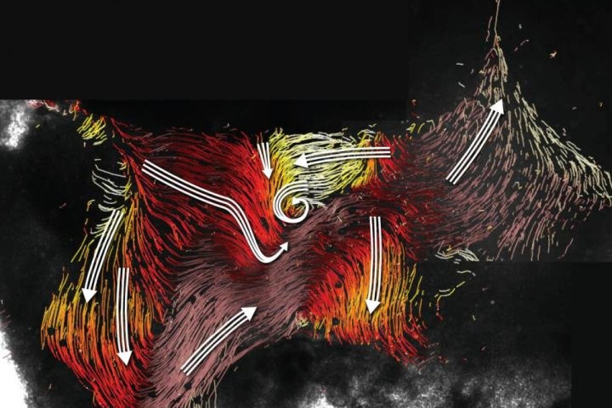
"In these images, we can see a complex network of fluid paths inside the cerebral ventricle. However, in contrast to the blood which flows through our blood vessels, these paths are not confined by walls. The exciting question for us was therefore: Is the flow pattern created solely by the synchronized beating of the cilia?" said Regina Faubel, first author of the study.
To find out, the researchers recorded the movement of the cilia and compared that to the flow of the fluorescent particles. They matched up, indicating that the cilia was indeed responsible for the movement of the fluid.
What particularly surprised the researchers was that the cilia didn't just beat in one direction as once believed, but they actually changed direction to a particular rhythm. They also found out that the movement of the cilia not only worked to deliver helpful chemicals to brain cells, but that the cilia could create counter flows to keep harmful substances away from parts of the brain.
"In the cerebral fluid of humans, there are hundreds — if not thousands — of physiologically active substances," Eichele said. "We are assuming that the network of flows we discovered plays an important role in distributing these substances. In other experiments, we would like to look at which messenger substances are transported via the flows, and where these are ultimately deposited in the tissue."
The group's work was published today in the journal Science.
Source: Max-Planck-Gesellschaft


