 One of the most common complications in implant dentistry is receding or very thin gum tissue which results in both aesthetic and functional problems. It is often due to poorly positioned dental implants with excessive inclination or tilt or inadequate bone and gum tissue. Patients with such complications report receding gum tissue, long implant crown, asymmetry, and pain related to peri-implantitis (inflammation around dental implants). But why does this happen and what can you do about it?
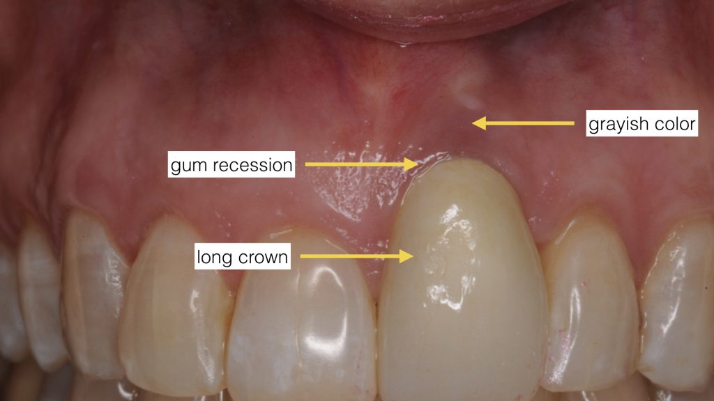
Proper dental implant position and sufficient bone and gum tissue are the critical factors in achieving success and prevention of such complications. If the treating dentist places the dental implant too far out or with excessive tilt, the gum tissue begins to recede. This happens even faster when the gum and bone tissues are already missing or very thin. The outcome is poor aesthetics with a long crown, gum recession, grayish gum tissue color, and poor accessibility for hygiene.
To prevent this complication the surgeon must place the implants in absolute correct position and angulation using digital dental implant diagnostics and planning. Also, proper bone and gum tissue must be present or augmented via grafting if inadequate. It all starts with simulation of the planned tooth and bite. A cone beam CT scan (CBCT) is obtained to assess bone quality and quantity. Then an optical scan of the patient’s teeth is obtained. Through a digital merging of these files and virtual simulation, the surgeon can then accurately plan and position the implant in proper angle while making sure the bone is adequate for its support. Finally the gum tissue is shaped through either a customized healing abutment or an implant-supported provisional restoration (prototype).
How about treatment for receding gum tissue around a poorly positioned dental implant? The only viable and predictable treatment is to atraumatically remove the dental implant using reverse torque. The surgeon will then regenerate the missing gum tissue and bone before replacement with a new dental implant in the right position using digital workflow in diagnostics and planning. The overall treatment may take as long as 9-12 months, but the result will be more aesthetic and stable for long term.
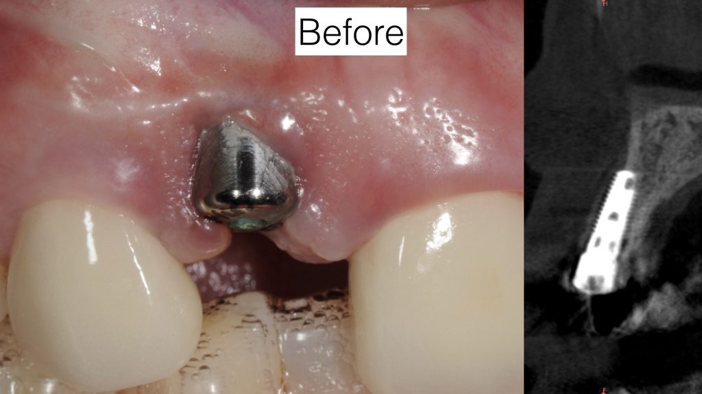
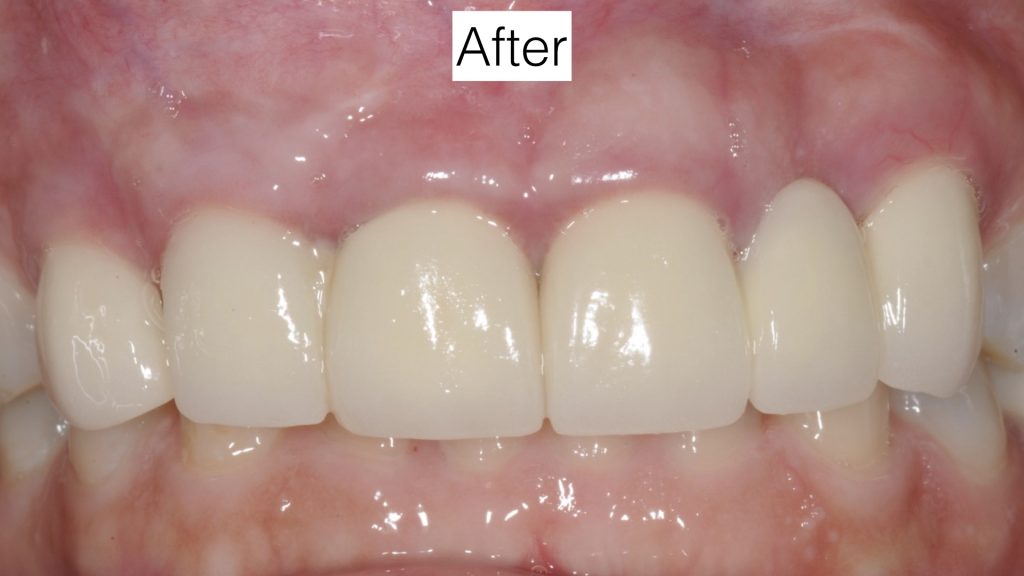
 This patient came to us 3 months after having dental implant placed by her dentist. She noticed a significant loss of gum tissue immediately after the procedure. The cone beam CT scan demonstrates excessive buccal position of the implant and inadequate bone and soft tissue coverage. The implant was subsequently removed at our office and the tissues were restored. A new implant in proper position was placed and restored with proper contours.
SEE FULL REPORT ON TREATMENT STRATEGY IN OUR PORTFOLIO SECTION
Dr. H. Ryan Kazemi is a board-certified oral and maxillofacial surgeon in Bethesda, MD.